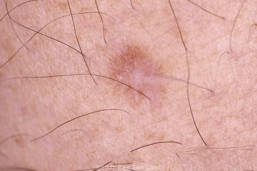
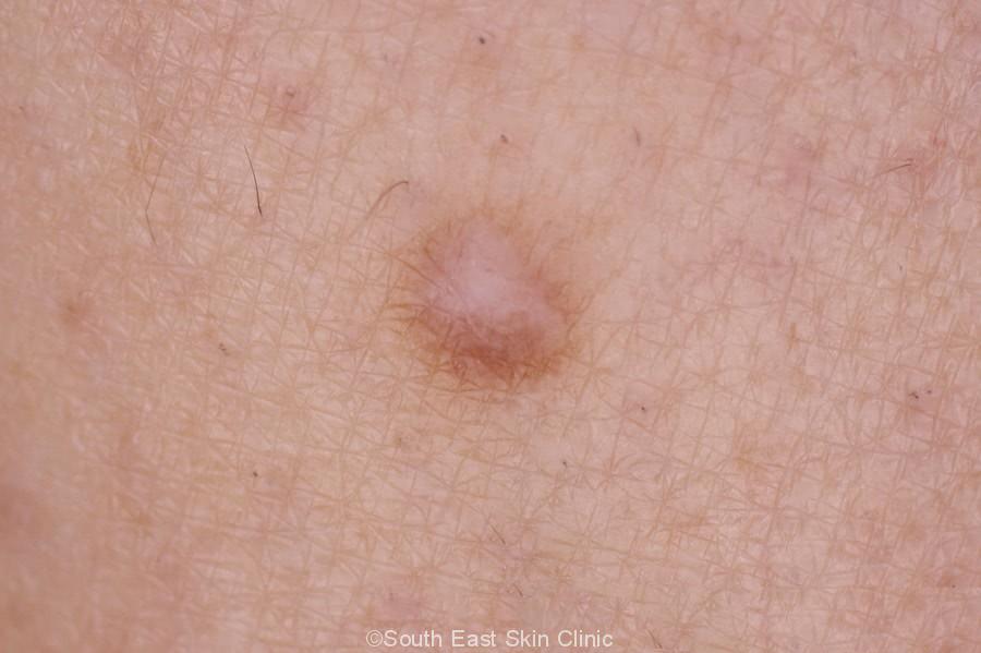
Dermatofibroma South East Skin Clinic blog
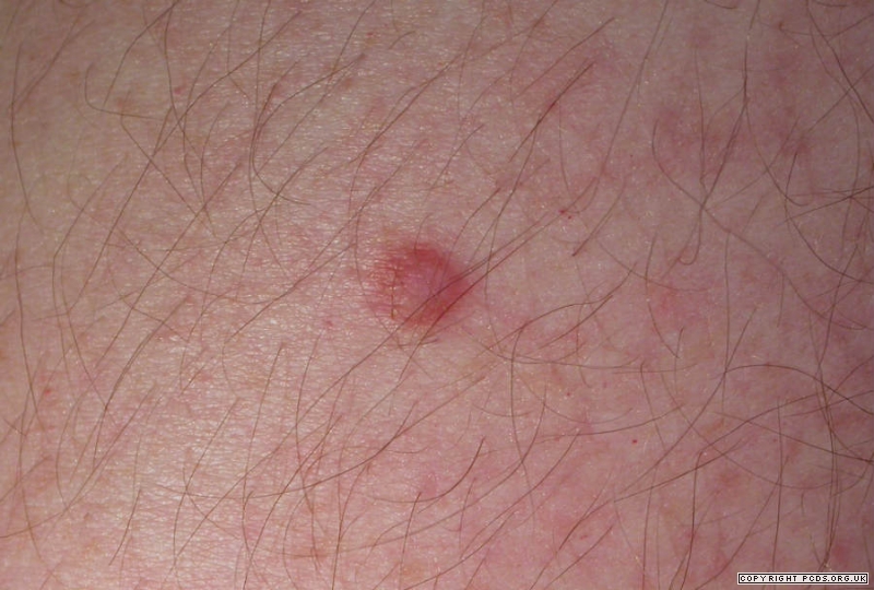
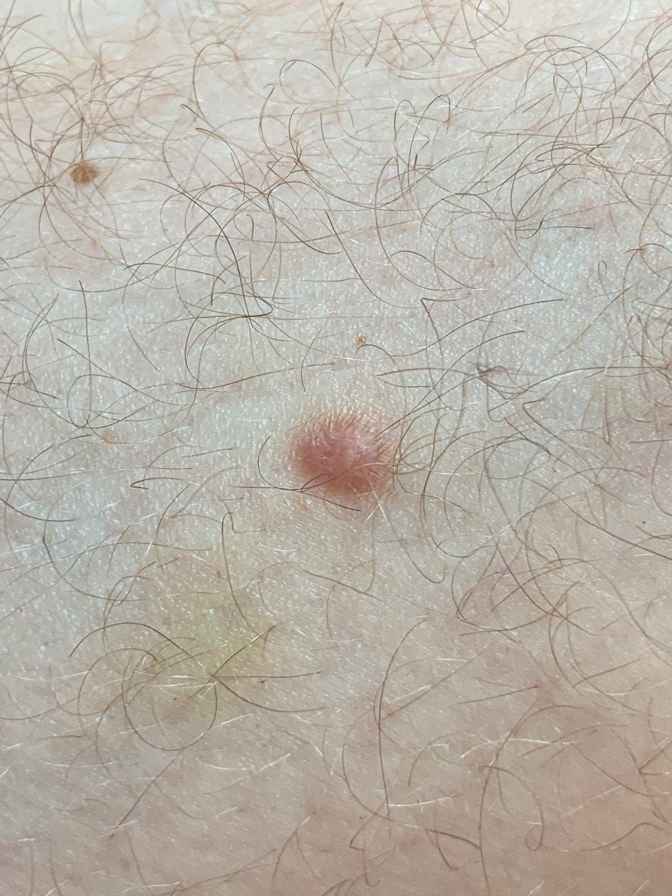
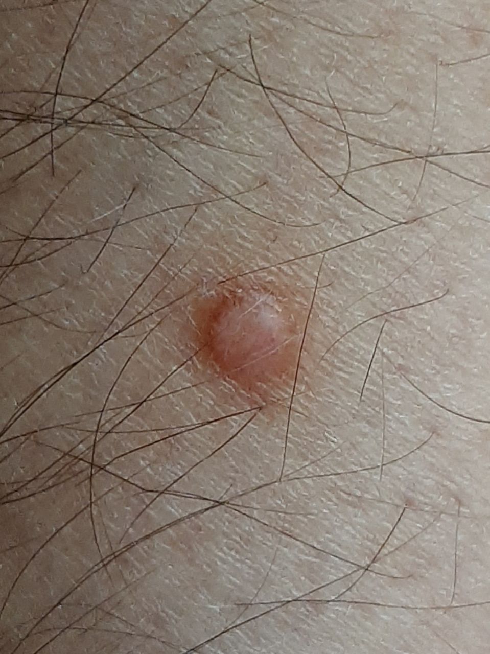
Dermatofibromas are firm, red-to-brown, small papules or nodules composed of fibroblastic tissue. They usually occur on the thighs or legs but can occur anywhere. Dermatofibroma Image courtesy of Marie Schreiner, PA-C. Dermatofibromas are common among adults, more so in women. Their cause is probably genetic.

O Que é Dermatofibroma e como é seu Tratamento
Il dermatofibroma è un tumore della pelle di natura benigna, piuttosto frequente. Questa neoformazione cutanea è costituita da una proliferazione di fibroblasti con localizzazione nel derma. Il dermatofibroma compare, di solito, in soggetti adulti, soprattutto di sesso femminile, tipicamente intorno ai 20-30 anni di età.
Dermatofibroma Didac Barco
Dermatofibroma è il termine medico che indica una categoria di tumori benigni della pelle, che originano dalle cellule dei tessuti connettivi fibrosi del derma. In genere, un dermatofibroma non rappresenta una condizione clinica pericolosa per l'essere umano; tuttavia la sua eventuale insorgenza richiede un adeguato e tempestivo consulto medico.
Dermatofibroma (ICD10 D23.9) Skinive Free AI Skin Diagnosis
Several variants of dermatofibroma have been described. They are essentially distinguished by their clinical and histopathological features. To review the mainfeaturesof these variants, a retrospective study of skin biopsies and tissue excisions of dermatofibromasperformed in the dermatology and venereology service at the Hospital Garcia de Orta between May 2007 and April 2012 was carried out.

Dermatofibroma Stock Image C049/8491 Science Photo Library
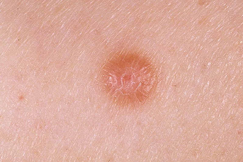
Dermatofibromas are small red-to-brown bumps that result from an accumulation of collagen, which is a protein made by the cells (fibroblasts) that populate the soft tissue under the skin. (See also Overview of Skin Growths .) Dermatofibromas are common among adults and usually appear as single firm bumps, often on the thighs or legs, and.
Dermatofibroma (ICD10 D23) Skinive AI
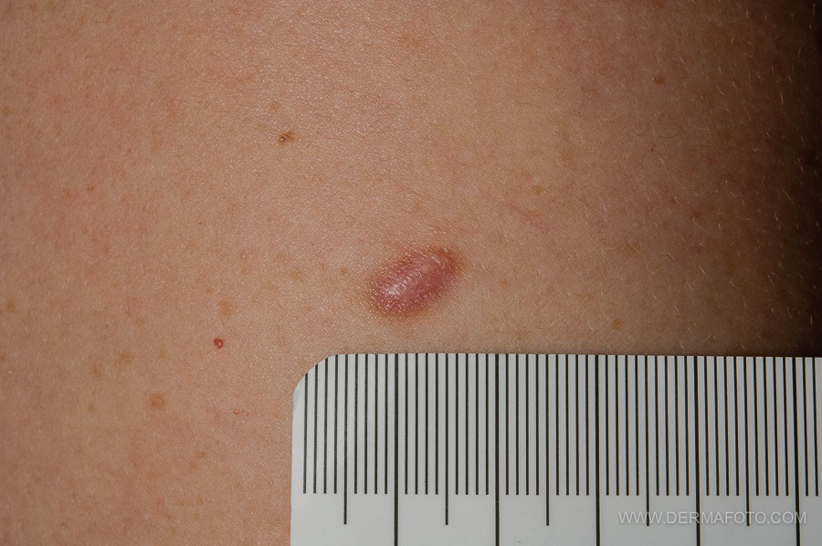
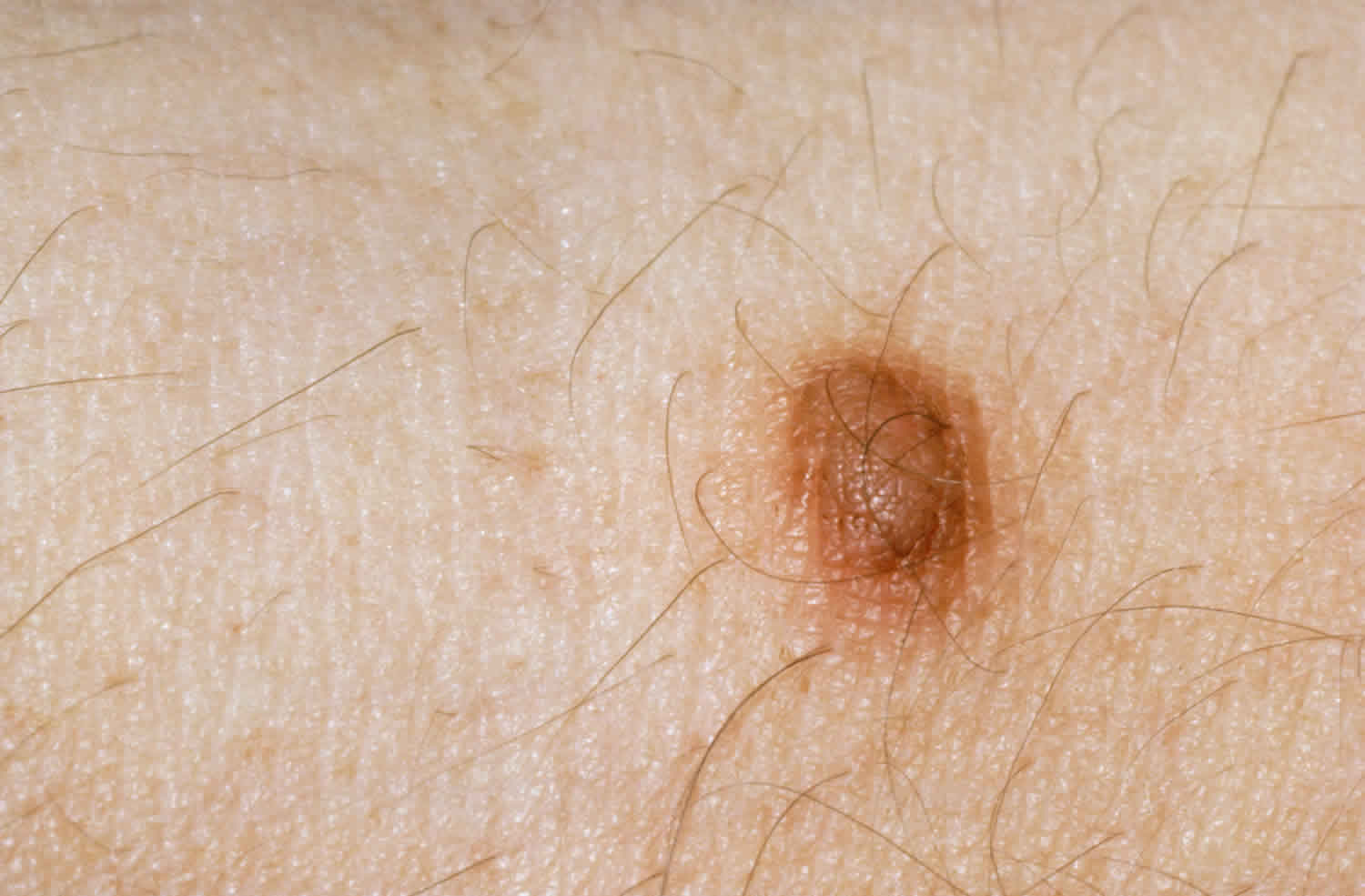
A dermatofibroma is a common benign fibrous nodule usually found on the skin of the lower legs. A dermatofibroma is also called a cutaneous fibrous histiocytoma. Who gets a dermatofibroma? Dermatofibromas are mostly seen in adults. People of every ethnicity can develop dermatofibromas.
Dermatofibroma South East Skin Clinic blog
Microscopic presentation of H&E-stained sections of an atrophic dermatofibroma on the right upper back of a 47-year-old man. Low (A) and higher (B) magnification of H&E-stained sections of an atrophic dermatofibroma shows a central depression (between blue arrows), epidermal acanthosis (thickening of the epidermis as shown between black bracket), basilar hyperpigmentation (yellow arrows), and.

Dermatofibroma Pictures, Removal, Treatment, Symptoms (2018 Updated)
A dermatofibroma is a nodule made of fibrous tissue. When a doctor squeezes the nodule during an examination, the overlying skin dimples. © DermNet New Zealand Causes and risk factors.

Dermatofibroma Dermatología BarcelonaDermatología Barcelona
What are dermatofibromas? Dermatofibromas are small, rounded noncancerous growths on the skin. The skin has different layers, including the subcutaneous fat cells, dermis, and epidermis. When.
dermatofibroma pictures pictures, photos
Results All our Moroccan patients had a dark skin phototype (Fitzpatrick scale types IV and V). A total of 14 morphological dermoscopic structures were distinguished, and 17 dermoscopic patterns were observed, with the most common pattern being the central white patch and peripheral pigment network (21%).
Dermatofibroma On Leg
How is a cellular dermatofibroma diagnosed? To diagnose a cellular dermatofibroma, your healthcare provider starts by looking at the lesion. You may have a skin biopsy to confirm if it's a dermatofibroma or another type of skin lesion. In a skin biopsy, your healthcare provider removes a small tissue sample. They send your tissue sample to a lab.
Dermatofibroma Pictures, Removal, Treatment, Symptoms (2018 Updated)
What Is It? Dermatofibromas are small, noncancerous (benign) skin growths that can develop anywhere on the body but most often appear on the lower legs, upper arms or upper back. These nodules are common in adults but are rare in children. They can be pink, gray, red or brown in color and may change color over the years.
Dermatofibroma South East Skin Clinic blog
Introduction. Dermatofibroma is a common benign tumour also known as fibrous histiocytoma. There is debate as to whether dermatofibroma has a reactive or neoplastic origin. The clinical lesion is a firm tan-brown nodule most commonly found on the legs. A number of histological variants exist.. Histology of dermatofibroma. Dermatofibromas are dermal tumours characterised by a poorly defined.
Dermatofibroma Pictures, Removal, Treatment, Causes, Symptoms, Images
Dermatofibroma (superficial benign fibrous histiocytoma) is a common cutaneous nodule of unknown etiology that occurs more often in women. Dermatofibroma frequently develops on the extremities.

Dermatofibroma Removal in Toronto Dermatofibroma Treatment
Dermatofibroma is a commonly occurring cutaneous entity usually centered within the skin's dermis. Dermatofibromas are referred to as benign fibrous histiocytomas of the skin, superficial/cutaneous benign fibrous histiocytomas, or common fibrous histiocytoma.

Dermatofibroma Benign Fibrous Histiocytoma... Academic Dermatology
Il dermatofibroma è un accumulo di collagene nel tessuto molle sotto la cute. La sua presenza è una condizione piuttosto comune, ma alcune persone possono sviluppare più dermatofibromi dislocati sul corpo. Dermatofibroma